AutoContour

AutoContour v2.7 unifies AutoContour and Limbus Contour into one AI-driven platform, delivering fast, consistent contouring with 480 guideline-based models across CT, MR, and CBCT. Works seamlessly with Eclipse and DICOM-compatible systems for flexible integration.

Schedule a demo
AutoContour
Automate your contouring
See AutoContour in action

Start Reviewing Automated Contours in Seconds

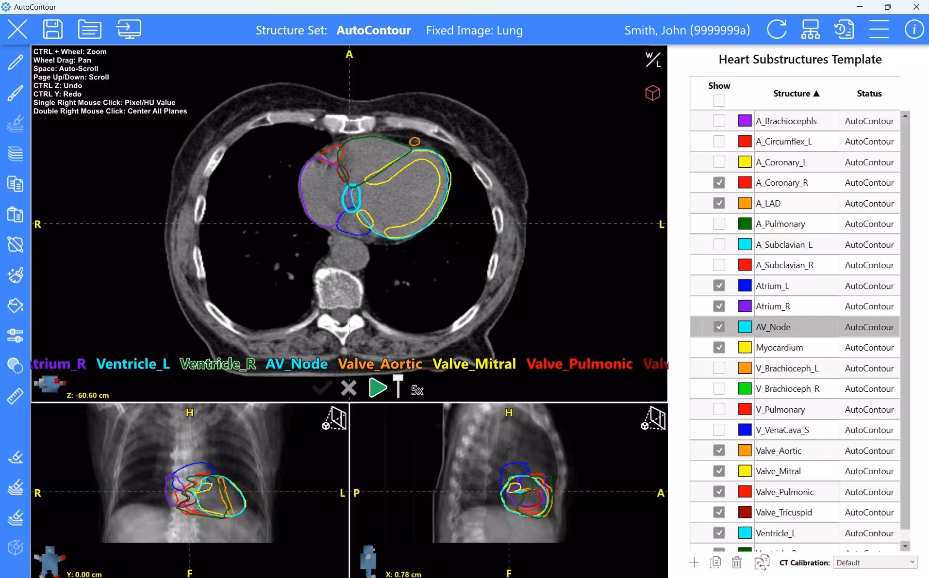
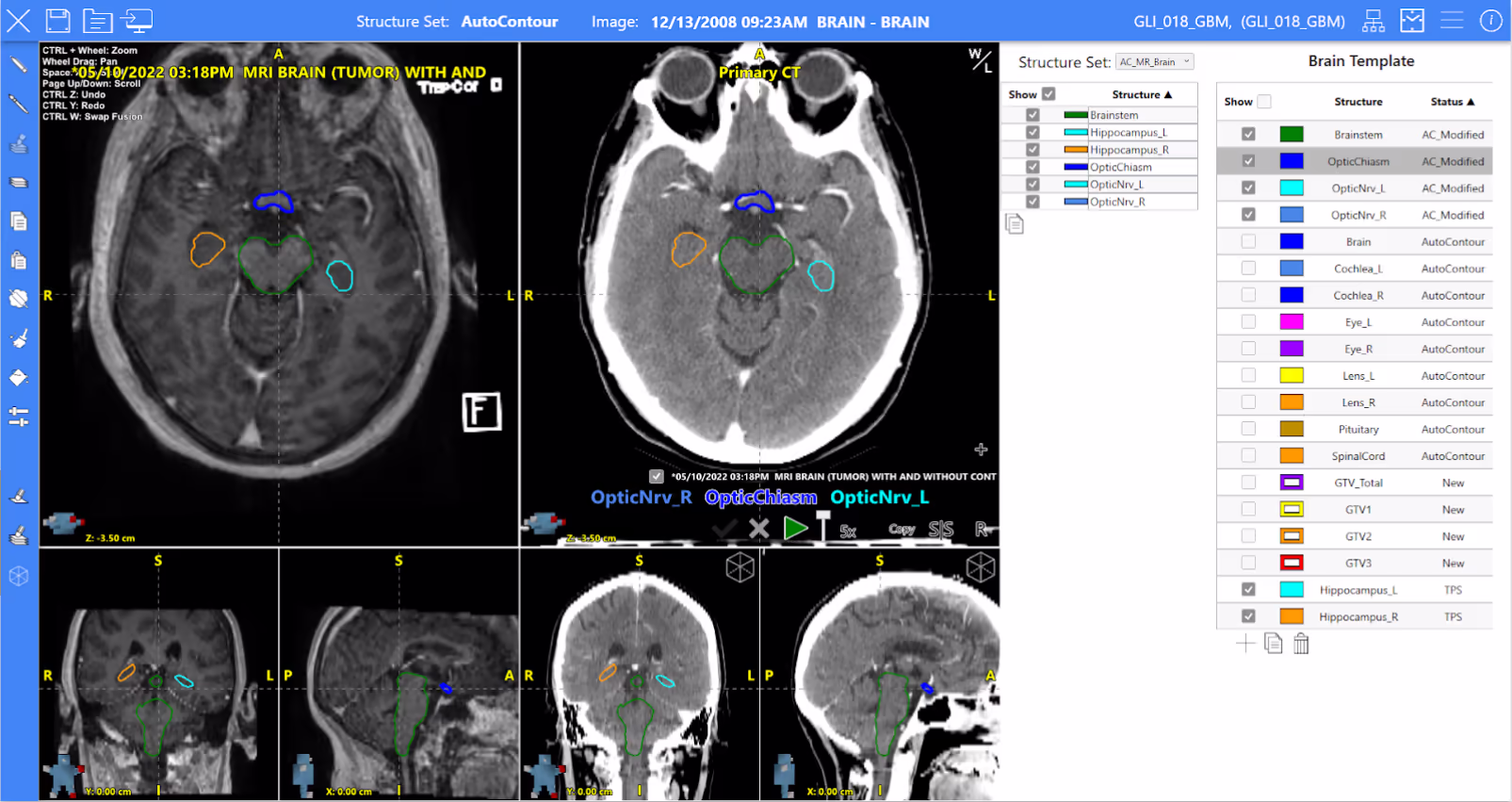
AutoContour v2.7 comes ready to use with 480 AI-trained models, including 115 lymph node models, across CT, MR, and CBCT. Built in alignment with international consensus guidelines, it delivers consistent, standardized contouring with planning-ready results in minutes, no setup required.

Head and Neck

Head and Neck

  • A_Carotid_L/R
  • Body+Mask
  • Bone_Hyoid
  • Bone_Mandible
  • Bone_Pterygoid_L/R
  • Bone_Skull
  • Bone_Teeth
  • Brain
  • Brainstem
  • BrachialPlex_L/R
  • BrachialPlexs
  • BuccalMucosa
  • Cavity_Oral
  • Cavity_Oral_Ext
  • Cerebellum
  • Clavicle_L/R
  • Cochlea_L/R
  • Cornea_2d_L/R
  • Cornea_L/R
  • CorpusCallosum
  • Cribriform
  • Dental_Artifact
  • Ear_Internal_L/R
  • Esophagus
  • Eye_L/R
  • Eyes
  • Falx
  • Glnd_Lacrimal_L/R
  • Glnd_Submand_L/R
  • Glnd_Thyroid
  • Hippocampus_L/R
  • InternalAuditoryCanal_L/R
  • Larynx
  • Larynx_Glottic
  • Larynx_NRG
  • Larynx_SG
  • Lens_L/R
  • Lips
  • Lobe_Temporal_L/R
  • Macula_L/R
  • Medulla
  • Midbrain
  • Musc_Constrict_2d
  • Musc_Constrict
  • Musc_Sclmast_L/R
  • OpticChiasm
  • OpticNrv_L/R
  • Optics
  • Parotid_Glnd_L/R
  • Parotid_L/R
  • Pharynx
  • Pituitary
  • Pituitary_3d
  • Pons
  • Retina_2d_L/R
  • Retina_L/R
  • Sinuses
  • Tentorium
  • Trachea
  • V_Jugular_L/R
  • Ventricle_Brain
Head and Neck Nodes

Head and Neck Nodes

  • LN_Neck_IA_2d
  • LN_Neck_IA_VI_2d
  • LN_Neck_II-IV_2d_L/R
  • LN_Neck_CerVII_2d_L/R
  • LN_Neck_2d_L/R
  • LN_Neck_VI_2d
  • LN_Neck_IA
  • LN_Neck_IB_2d_L/R
  • LN_Neck_IB_L/R
  • LN_Neck_IB-V_L/R
  • LN_Neck_II_2d_L/R
  • LN_Neck_II_L/R
  • LN_Neck_II-V_L/R
  • LN_Neck_III_2d_L/R
  • LN_Neck_III_L/R
  • LN_Neck_IV_2d_L/R
  • LN_Neck_IV_L/R
  • LN_Neck_II-IV_L/R
  • LN_Neck_V_2d_L/R
  • LN_Neck_V_L/R
  • LN_Neck_VIA
  • LN_Neck_VIIA_2d_L/R
  • LN_Neck_VIIA_L/R
  • LN_Neck_VII_2d_L/R
  • LN_Neck_VIIB_2d_L/R
  • LN_Neck_VIIB_L/R
  • LN_Post_Neck_L/R
Breast Nodes

Breast Nodes

  • LN_Ax_L1_ESTRO_2d_L/R
  • LN_Ax_L2_ESTRO_2d_L/R
  • LN_Ax_L2_InPec_ESTRO_2d_L/R
  • LN_Ax_L3_ESTRO_2d_L/R
  • LN_IMN_ESTRO_2d_L/R
  • LN_IMN_Expand_ESTRO_2d_L/R
  • LN_InPec_ESTRO_2d_L/R
  • LN_Sclav_ESTRO_2d_L/R
  • LN_Ax_L/R
  • LN_Ax_L1_2d_L/R
  • LN_Ax_L1_ESTRO_L/R
  • LN_Ax_L1_L/R
  • LN_Ax_L2_2d_L/R
  • LN_Ax_L2_ESTRO_L/R
  • LN_Ax_L2_L/R
  • LN_Ax_L2_L3_L/R
  • LN_Ax_L3_2d_L/R
  • LN_Ax_L3_ESTRO_L/R
  • LN_Ax_L3_L/R
  • LN_Ax_Sclav_2d_L/R
  • LN_IMN_2d_L/R
  • LN_IMN_Expand_2d_L/R
  • LN_IMN_L/R
  • LN_IMN_RC_L/R
  • LN_InPec_ESTRO_L/R
  • LN_Sclav_2d_L/R
  • LN_Sclav_ESTRO_L/R
  • LN_Sclav_L/R
  • LN_Sclav_RC_L/R
Breast and Chestwall

Breast and Chestwall

  • A_Aorta
  • A_Aorta_Asc
  • A_Aorta_Dsc
  • A_LAD
  • A_Pulmonary
  • Atrium_L/R
  • BrachialPlex_L/R
  • BrachialPlexs
  • Breast_Implant_L/R
  • Breast_L/R
  • Breast_Prone
  • Breast_RTOG_L/R
  • Breast_Wire_L/R
  • Breasts_RTOG
  • Chestwall_ESTRO_L/R
  • Chestwall_L/R
  • Chestwall_RC_L/R
  • Clavicle_L/R
  • Esophagus
  • Glnd_Thyroid
  • Heart
  • Heart_Prone
  • Humerus_L/R
  • Liver
  • Lung_L/R
  • Lungs
  • Nipple_L/R
  • Nipple_Prone
  • Pericardium
  • Stomach
  • V_Venacava_I
  • V_Venacava_S
  • Ventricle_L/R
Thorax

Thorax

  • A_Aorta
  • A_Aorta_Asc
  • A_Aorta_Dsc
  • A_Brachiocephls
  • A_Circumflex_L
  • A_Coronary_2d_R
  • A_Coronary_L
  • A_Coronary_R
  • A_LAD
  • A_Pulmonary
  • A_Subclavian_L/R
  • Atrium_L/R
  • AV_Node
  • Bone_Sternum
  • BrachialPlex_L/R
  • BrachialPlexs
  • Bronchus
  • Carina
  • Chestwall_Anat
  • Chestwall_Anat_L/R
  • Chestwall_2cm_L/R
  • Chestwall_OAR
  • Clavicle_L/R
  • Esophagus
  • GreatVessels
  • Heart
  • Heart_Prone
  • Heart+A_Pulm
  • Lung_L/R
  • Lungs
  • Musc_PecMinor_L/R
  • Myocardium
  • Pericardium
  • Pericardium_Inf
  • Pericardium_Inf+A_Pulm
  • Trachea
  • V_Brachioceph_L/R
  • V_Pulmonary
  • V_Venacava_I
  • V_Venacava_I_UKSABR
  • V_Venacava_S
  • V_Venacava_S_UKSABR
  • Valve_Aortic
  • Valve_Mitral
  • Valve_Pulmonic
  • Valve_Tricuspid
  • Ventricle_L/R
Abdomen

Abdomen

  • A_Aorta
  • A_Aorta_Dsc
  • A_Celiac
  • A_Mesenteric_S
  • BileDuct_Common
  • Bowel
  • Bowel_Bag
  • Bowel_Large
  • Bowel_Small
  • Duodenum
  • Esophagus
  • Gallbladder
  • Kidney_L/R
  • Kidney_Outer_L/R
  • Kidneys
  • Kidneys_Outer
  • Liver
  • LN_Paraaortic
  • Pancreas
  • Spleen
  • Stomach
  • V_Venacava_I
  • V_Venacava_I_UKSABR
Male Pelvis

Male Pelvis

  • A_Aorta
  • A_Aorta_Dsc
  • Barrigel
  • Bladder
  • Bone_Ilium
  • Bone_Ilium_L/R
  • Bone_Ischium_L/R
  • Bone_Pelvic
  • Bone_Sacrum
  • Bowel
  • Bowel_Bag
  • Bowel_Large
  • Bowel_Small
  • Canal_Anal
  • Colon_Sigmoid
  • Femur_Head_L/R
  • Femur_L/R
  • Femur_RTOG_L/R
  • Femurs_RTOG
  • Genitals_M
  • Iliac_Int_L/R
  • Iliac_L/R
  • LN_Inguinofem_L/R
  • LN_Mesorectum
  • LN_Paraaortic
  • LN_Pelvics
  • LN_Pelvics_NRG
  • LN_Presacral
  • Marrow_Ilium_L/R
  • Marrow_Pelvis
  • Musc_Iliopsoas_L/R
  • PenileBulb
  • Prostate
  • Prostate-Spacergel_VUE
  • Prostate+SeminalVes
  • Prostate+SV-Spacergel_VUE
  • ProstateBed
  • ProstateFiducials
  • Prostate-Gel_Fiducials
  • Bone_PubicSymphys
  • Rectum
  • Rectum-Spacergel_VUE
  • SpaceOARVue
  • SacralPlex_L/R
  • SeminalVes
  • V_Venacava_I
  • Vessels_Pelvis
Female Pelvis

Female Pelvis

  • A_Aorta
  • A_Aorta_Dsc
  • Bladder_F
  • Bone_Ilium
  • Bone_Ilium_L/R
  • Bone_Ischium_L/R
  • Bone_Pelvic
  • Bone_Sacrum
  • Bowel_Bag_F
  • Bowel_F
  • Bowel_Large_F
  • Bowel_Small_F
  • Canal_Anal_F
  • Colon_Sigmoid
  • Femur_Head_L/R
  • Femur_L/R
  • Femur_RTOG_L/R
  • Femurs_RTOG
  • Genitals_F
  • Iliac_Int_L/R
  • Iliac_L/R
  • LN_Pelvics_F
  • LN_Presacral
  • LN_Inguinofem_L/R
  • LN_Paraaortic
  • LN_Mesorectum
  • Marrow_Ilium_L/R
  • Marrow_Pelvis
  • Musc_Iliopsoas_L/R
  • Parametrium
  • Bone_PubicSymphys
  • Rectum_F
  • SacralPlex_L/R
  • UteroCervix
  • Vagina
  • V_Venacava_I
  • Vessels_Pelvis
HDR Female Pelvis

HDR Female Pelvis

  • HDR_Bladder
  • HDR_Bowel
  • HDR_Canal_Anal
  • HDR_Cylinder
  • HDR_Rectum
  • HDR_Ring
  • HDR_Colon_Sigmoid
  • HDR_Urethra
  • Foley_Balloon
Brain MRT1

Brain MRT1

  • Amygdala_L/R
  • Brain
  • Brainstem
  • Cerebellum
  • Cornea_L/R
  • CorpusCallosum
  • Eye_L/R
  • Falx
  • Hippocampus_L/R
  • Hypo_True
  • Hypothalamus
  • Lens_L/R
  • Medulla
  • Midbrain
  • OpticChiasm
  • OpticNrv_L/R
  • Optics
  • OpticTract_L/R
  • Pituitary
  • Pons
  • Retina_L/R
  • Sinuses
  • SpinalCord_Cerv
  • Tentorium
  • Thalamus
  • Ventricle_Brain
Male Pelvis MRT2

Male Pelvis MRT2

  • A_Pud_Int_L/R
  • Bladder
  • Bladder_TRUFI
  • Bladder_Trigone
  • Colon_Sigmoid
  • External_Pelvis
  • Femur_L/R
  • Glnd_Prostate
  • NVB_L/R
  • PenileBulb
  • PenileBulb_TRUFI
  • Prostate
  • PubicSymphys
  • Rectal_Spacer
  • Rectum
  • Bone_Sacrum
  • SeminalVes
  • Urethra
HDR Female Pelvis MRT2

HDR Female Pelvis MRT2

  • HDR_Bladder
  • HDR_Bowel
  • HDR_Canal_Anal
  • HDR_Colon_Sigmoid
  • HDR_Rec-Canal
  • HDR_Rectum
  • HDR_Urethra
Male Pelvis CBCT

Male Pelvis CBCT

  • Bladder_CBCT
  • Femur_L/R_CBCT
  • LN_Pelvics_CBCT
  • Prostate_CBCT
  • Rectum_CBCT
  • SeminalVes_CBCT
MultiSite

MultiSite

  • Body
  • CaudaEquina
  • External
  • Rib
  • Rib_L/R
  • Rib01_L/R
  • Rib02_L/R
  • Rib03_L/R
  • Rib04_L/R
  • Rib05_L/R
  • Rib06_L/R
  • Rib07_L/R
  • Rib08_L/R
  • Rib09_L/R
  • Rib10_L/R
  • Rib11_L/R
  • Rib12_L/R
  • Skin
  • SpinalCanal
  • SpinalCord
  • VB
  • VB_C1
  • VB_C2
  • VB_C3
  • VB_C4
  • VB_C5
  • VB_C6
  • VB_C7
  • VB_L1
  • VB_L2
  • VB_L3
  • VB_L4
  • VB_L5
  • VB_T01
  • VB_T02
  • VB_T03
  • VB_T04
  • VB_T05
  • VB_T06
  • VB_T07
  • VB_T08
  • VB_T09
  • VB_T10
  • VB_T11
  • VB_T12
AC
AutoContour models are developed in accordance with established international contouring guidelines, including RTOG, NRG, RADCOMP, ESTRO, DAHANCA, EORTC, GORTEC, TROG, HKNPCSG, NCIC CTG, NCR, and more. Combined with AAPM TG-263 nomenclature, this ensures consistent, high-quality contours across clinicians, sites, and clinical workflows.
Request the AutoContour Model Atlas

Image Registration Workspace

Rigid Registration

  • Automatic and Manual Editing
  • Shared Frame of Reference Display
    • Compatible modalities:
      CT, CBCT, MR, CT-PET
  • 4D-CT Handling
    • Shared Frame of Reference Display
    • ITV/PRV contour propagation
  • Structure Transfer
  • Structure Analysis Tools
  • DICOM and ESAPI Compatible

Deformable Registration

  • Evaluation Tools
    • Deformed structure analysis
    • Deformed grid/heat map
    • Deformed Dose display
  • Deformed Structure Transfer
  • Export Deformed Registration
  • Deform Dose Review Workspace Improvements NEW!

Re-irradiation Workflows / Adaptive Plan Sums

  • Multi-modality support across CT, MR, and CBCT
  • Deformable registration for accurate anatomical mapping
  • Track anatomical change across timepoints
  • Evaluate cumulative dose to support re-irradiation decisions
  • Integrated registration and dose workflows in a single platform
  • Accumulate dose across prior and current treatments *
  • EQD2/BED evaluation available *

* When used with ClearCheck

“AutoContour’s deformed sum provides a more accurate representation during treatment courses requiring adaptive planning, such as head and neck cases, given significant changes in anatomy.”
Dr Alexander Diaz, Radiation Oncologist, Murray-Calloway County Hospital

High-Quality Contours in a Fraction of the Time

97.4%

Time Savings *

AutoContour vs Manual Contouring Time Savings - AutoContour Saved 1400 RTT hours

1400

RTT Hours Saved *

* Modeled across network for first 8 months of use

Planning Structure Generation

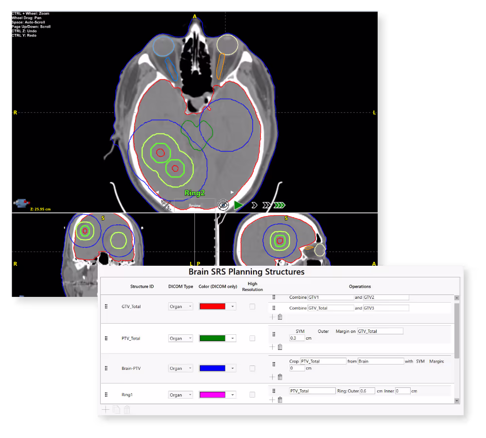
AutoContour automatically generates optimization structures in seconds, eliminating manual setup and improving consistency across patients.

  • Advanced structure creation tools for efficient planning workflows
  • Automatic generation of margins, rings, overlaps, and cropped structures
  • Boolean operations for precise structure combination and modification
  • Template-driven workflows to standardize structure creation
  • Configure once, apply across patients for scalable consistency
  • Import user and global planning structure templates for flexibility

Streamlined Editing and Review

  • Single-click grouped review with automatic plane alignment and synchronized scrolling
  • Configurable viewing controls with structure-specific scroll speed, window/level, and zoom
  • Efficient slice management with “keep every Nth slice” for faster refinement
  • Comprehensive editing tools including pencil, adaptive brush, interpolation, and multi-slice editing
  • Edit Assist Tool predicts contour propagation across slices to accelerate delineation
  • Dedicated Eraser Mode appears automatically with the brush tool for precise corrections NEW!

Vendor Neutral Contouring Solution

Seamlessly integrate with Eclipse (read/write), or use standalone DICOM capability with any treatment planning system, leveraging AI deep learning on both cloud based or local resources.

Eclipse Workflow

Integration with Eclipse & ClearCheck

CT Scanner

CT or MR Scanner

Send images to Eclipse

Right Arrow
Eclipse

Eclipse

Launch AutoContour via Eclipse API

Right and Left Arrows
AutoContour

AutoContour

Automatically contour structures and write back to Eclipse

DICOM Workflow

Vendor Neutral for TPS Flexibility

CT Scanner

CT or MR Scanner

Send images to AutoContour

Right Arrow
AutoContour

AutoContour

Automatically contour structures, review, and export

Right Arrow
TPS

Treatment Planning

Import structures and start planning process

Zero-click Workflow

Clinic Supported Review Process

CT Scanner

CT or MR Scanner

AutoContour listens for new DICOM data

Right Arrow
AutoContour

AutoContour

Processes structures; upon completion, automatically exported

Right Arrow
TPS

Treatment Planning

Import structures and start planning process

“Several features stand out across Radformation’s suite of tools. The brachial plexus contouring in AutoContour has been particularly valuable for ensuring accurate and efficient delineation.”

- Tosin Kayode, Director of Medical Dosimetry, Winship Cancer Institute.

“My everyday essentials are AutoContour, ClearCheck, and ClearCalc with RadMonteCarlo - they're absolutely central to how I work.”

- Michael Trager, Senior Radiation Physicist, Northwell Health

“With the volume of patients we treat daily, AutoContour significantly cuts down contouring time and improves consistency across patients”

- Nusirat Adeddewe, Medical Physicist, Medserve-Luth Cancer Center

“AutoContour is an intuitive and practical solution that was rapidly adopted due to its ease of use. It requires minimal post-processing, significantly reducing the time spent on manual contouring. Furthermore, the technology facilitates remote support for other units, ensuring a consistent standard of contouring across the network - even in high-demand or understaffed environments.”

- Rinette Koenig, Radiation Therapist, Icon Oncology, South Africa

    AutoContour Overview

    Generate consistent, high-quality OAR and planning structure contours in seconds, reducing manual effort and accelerating time to plan.

    Keep up with Radformation

    Get the latest news, announcements, and product updates delivered straight to your inbox.